Structures nobody could have guessed
Diffraction hands you intensities and keeps the phases, so a structure is not something you invert a pattern to get. Dorothy Hodgkin solved penicillin against the settled opinion of the most eminent chemists in Britain, then vitamin B₁₂ with 181 atoms and no prior knowledge at all, then insulin after thirty-four years.
- State the phase problem quantitatively, and say why it cannot be brute-forced
- Explain how a heavy atom supplies phases the experiment never measured
- Describe how the structure of penicillin was settled against expert chemical opinion
- Say what changed between penicillin in 1945 and vitamin B₁₂ in 1956
Bragg handed chemistry an instrument, and the instrument came with a hole in it. Every spot is a complex number. The structure factor F has a magnitude and a phase. Get both, transform them, and the electron density appears with the atoms in it. That step is exact and it is not the hard part. The detector records |F|², and nothing else. Intensity is a photon count. It gives you the magnitude squared and it gives you not the faintest indication of the phase. No instrument has ever measured one directly. So the calculation needs two numbers per reflection, the experiment supplies one, and the one that is missing turns out to carry more of the structural information than the one you have. What this meant in practice, through the 1920s and 1930s, is that you could solve a structure only if you already very nearly knew the answer. You proposed an arrangement, calculated what its pattern would be, compared it with the observed intensities, and adjusted. That works for rock salt, where there are two candidates and one is silly. It works for hexamethylbenzene, where the chemistry has narrowed things to a ring. It does not work for a molecule nobody has a proposal for.
Start with the true phases and note that the peaks land on the atoms. Then drag the phase-error slider to the right and watch the amplitude bars, which do not move.
The size of the easiest case
A centrosymmetric projection of a small molecule yields 100 independent reflections. Every structure factor is real, so each phase is simply a sign — plus or minus. How many distinct combinations of signs are there?
- Independent reflections
- n = 100
- Possible phases per reflection
- 2 (+ or −)
- Reflections are independent
- each sign is free of the others
Dorothy Mary Crowfoot was born in Cairo in 1910, read chemistry at Somerville College, Oxford, and went to Cambridge in 1932 to work with J. D. Bernal. In 1934 the two of them took the first X-ray diffraction photograph of a protein, from a crystal of pepsin — and the reason it worked is worth knowing, because it is a very small piece of technique with an enormous consequence. Everyone before them had mounted crystals dry. Protein crystals are around half water, and drying one destroys the order that makes it a crystal. Bernal and Crowfoot sealed theirs in a thin glass tube along with the liquid it had grown in, and the spots appeared. That photograph is the moment it becomes conceivable that the structure of a protein might one day be known. The same year, aged 24, she was diagnosed with rheumatoid arthritis. It deformed her hands progressively for the rest of her life, and she worked with it for sixty years — the last of them from a wheelchair, still travelling.
This is the same picture with the phases taken from the heavy atom alone. Compare the map with the true atom positions marked below it — then press "Light atoms only" and watch what happens without a rubidium in the cell.
The X-ray structure of penicillin
- The question
- Penicillin was saving lives and nobody knew what it was. Two structures had been proposed for it: a β-lactam, with a strained four-membered nitrogen-containing ring fused to a five-membered thiazolidine, and a tricyclic oxazolone–thiazolidine arrangement using only ordinary five-membered rings. Which is it?
- The apparatus
- Crystalline salts of benzylpenicillin — sodium, potassium and rubidium — grown so that the same molecule could be examined in three different lattices. Rubidium, with 36 electrons against the 23 non-hydrogen atoms of the penicillin itself, is the heavy atom. Two-dimensional Fourier projections computed by hand and on Hollerith punched-card machines, at a time when a single map was weeks of arithmetic.
On the chemical evidence and on the judgement of the senior structural chemists involved — Robert Robinson foremost among them — the oxazolone structure. Four-membered rings are strained, rare, and were widely regarded as too unstable to be sitting at the centre of a usable drug.
The β-lactam. A four-membered ring containing nitrogen, fused to the thiazolidine, exactly as Chain had proposed and as the chemists had rejected.
How sure could they be? The argument rests on the three salts agreeing. The same molecular geometry emerging from three different crystal lattices is very hard to explain as an artefact of any one of them — and the rubidium salt supplied phases the other two could not.
A measurement settled a structural question against the considered opinion of the most eminent chemists in Britain, and it settled it in a way anyone could check by growing the same crystals. That is worth stating plainly because it is the entire claim this site makes. It also mattered enormously in practice: the β-lactam ring is the part of penicillin that does the work, and knowing it was there is what made it possible to strip the rest away and rebuild.
It is easy to tell this as a story about a stubborn establishment and a lone woman who was right. That is not quite what happened, and the real version is more interesting. Robert Robinson was Waynflete Professor of Chemistry at Oxford and would win the 1947 Nobel Prize in Chemistry. He was, by a wide margin, one of the best structural chemists alive, and his intuition about what molecules are like had been right for thirty years. He thought the β-lactam was wrong. His reasoning was good. Four-membered rings are strained. They are rare in nature. They are unstable — which is exactly the objection: a molecule you can put in a bottle and inject into people should not have one at its centre. Against that, the oxazolone structure uses only comfortable five-membered rings and accounts for most of the chemical behaviour. Ernst Chain, who had proposed the β-lactam, was a biochemist arguing about structure with organic chemists, in a subject where they had the authority. What resolved it was not a better argument. Robinson could not have been talked out of his position and should not have been. What resolved it is that Hodgkin grew three salts, computed the maps, and the four-membered ring was in all three of them. The instability the chemists objected to is real. It is also the mechanism: the ring is strained enough to open when it meets the enzyme a bacterium uses to cross-link its cell wall, and it destroys the enzyme in the process. Robinson’s objection was correct chemistry pointing at the wrong conclusion, which is a much more common failure than being simply mistaken.
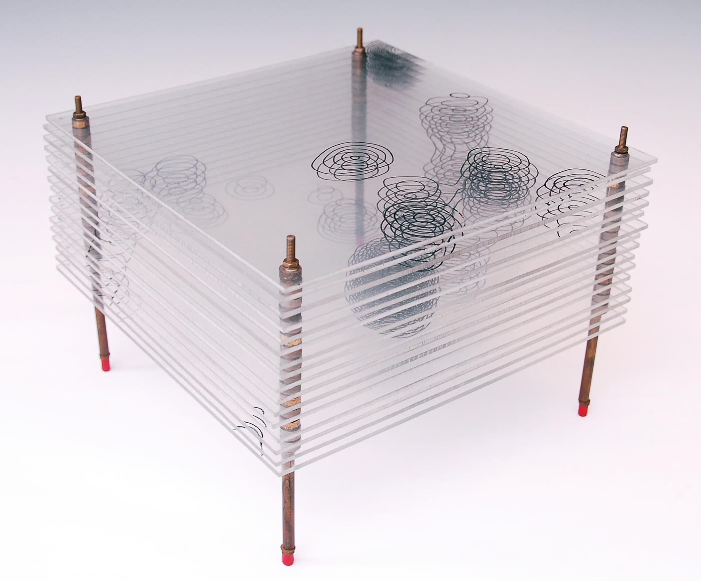
Museum of the History of Science, University of Oxford, 2013-10-15 14:20:26. CC BY-SA 3.0
The structure of vitamin B₁₂
- The question
- Pernicious anaemia had been treatable with raw liver since 1926 and nobody knew what the active substance was. B₁₂ was crystallised in 1948. What is it? Chemical degradation had failed: the fragments could not be reassembled into anything consistent.
- The apparatus
- Crystals of the vitamin and of a hexacarboxylic acid derived from it. The molecule supplies its own heavy atom — a cobalt sitting at the centre — which is why B₁₂ was solvable at all. Thousands of reflections, and Fourier syntheses far beyond hand computation: the calculations were run on the SWAC, one of the earliest stored-program electronic computers, by Kenneth Trueblood in Los Angeles, with data and results crossing the Atlantic by post.
Nothing specific. This is the point — there was no candidate structure to test. Chemistry had produced fragments and no way to put them together, and the molecule was far larger than anything previously solved from scratch.
A corrin ring — related to the porphyrin of haem but with one bridging carbon missing — with cobalt at its centre, a nucleotide loop tucked underneath, and 181 atoms in all, 93 of them heavier than hydrogen. Including a cobalt–carbon bond, the first ever found in a biological molecule.
How sure could they be? The corrin ring is not a porphyrin, and the difference is a single missing carbon in one bridge. Nothing in the chemical evidence had suggested it, and no chemist proposed it. It came out of the maps.
This is the demonstration that crystallography can determine a structure with no prior structural knowledge whatever — that it is a method of discovery and not merely of confirmation. Lawrence Bragg, who had solved rock salt with two positions to choose between, said solving B₁₂ was as difficult as breaking an enemy cipher. It also converted the technique into something the whole of biochemistry would come to depend on.
And then insulin, which she began in 1935 and finished in 1969. Thirty-four years. Fifty-one amino acids in two chains, several hundred non-hydrogen atoms, no convenient heavy atom of its own, and crystals with a great deal of water in them. She obtained her first insulin diffraction photographs as a young researcher, put the problem down when it proved impossible, and picked it up again repeatedly across four decades while solving penicillin and B₁₂ in between. She was awarded the Nobel Prize in Chemistry in 1964 — for penicillin and B₁₂. Insulin was still unsolved. It came out five years after the prize. It is worth pausing on what that means about the shape of a scientific life. The public record is three structures and a medal. The actual work is thirty-four years of a problem that would not yield, carried alongside everything else, with no guarantee at any point that it ever would.
The computers solved these structures — vitamin B₁₂ came out of one of the first electronic machines.
The SWAC did the Fourier syntheses, and without it B₁₂ would have taken decades rather than years. But a Fourier synthesis is arithmetic, and arithmetic was never the obstacle: the obstacle was that the phases going into it were not measured and had to be supplied. Every phase the machine used came from a decision somebody made — locate the cobalt in a Patterson map, assume its phases, read the resulting map, judge by chemistry which peaks are real atoms and which are the mirror ghost, add them, recompute. The machine could not do any of that and was never asked to. It is a fair description of B₁₂ to say that a computer did in weeks what a person could not do in a lifetime, and a false one to say that a computer solved it. The same distinction is worth carrying into any modern claim of the same shape.
I was captured for life by chemistry and by crystals.
- 1934Bernal and Crowfoot photograph a wet pepsin crystal: the first diffraction pattern from a protein.
- 1934Patterson publishes the map that gives interatomic vectors from intensities alone — no phases needed.
- 1934She is diagnosed with rheumatoid arthritis, aged 24.
- 1935First X-ray photographs of insulin crystals. The structure will take another 34 years.
- 1942Work on penicillin begins, in wartime secrecy.
- 1945The β-lactam ring, against the settled opinion of the chemists. Publication waits until 1949.
- 1948Vitamin B₁₂ is crystallised. Nobody has any idea what it is.
- 1956B₁₂ solved: 181 atoms, a corrin ring, and a cobalt–carbon bond nobody expected.
- 1957Beecham isolate 6-APA and begin building semi-synthetic penicillins on Hodgkin’s ring.
- 1964Nobel Prize in Chemistry. "Oxford housewife wins Nobel."
- 1969Insulin, at last — five years after the prize she was given for the other two.